Jingle Bells, Your Butt Smells: A Surgeon’s Protocol for the BBL Recovery Nobody Talks About

Jingle Bells, Your Butt Smells: A Surgeon's Protocol for the BBL Recovery Nobody Talks About. Dr. Frank Agullo, MD, FACS, double board-certified plastic surgeon at Southwest Plastic Surgery in El Paso, Texas.

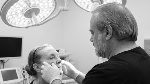
Australia’s GP-trade journal, Medical Republic, opened a piece on me last month with the line above, and I will admit to laughing out loud when the alert hit my phone. The piece, titled “Jingle bells, your butt smells,” reprinted the four-bullet post-operative hygiene protocol I wrote for Brazilian Butt Lift patients, credited me as a founding vice-president of the World Association of Gluteal Surgeons, and pushed the conversation out of the back rooms of the practice and into the GP literature halfway around the world.

The reason it traveled is that the topic genuinely is one of the least discussed parts of BBL recovery. The reason I wrote the protocol down in the first place is that nearly every BBL patient in my practice eventually asks me a quieter version of the same question. So here is the longer surgeon-to-surgeon version of the protocol, with the clinical reasoning behind each step.

What the Inside of a Fresh BBL Looks Like at Week Two

A BBL is two operations done together. A liposuction harvest from the donor sites, which can include the abdomen, the flanks, the back, the lower back, the inner thighs, and any other compartment from which the fat has been planned. And a structured gluteal injection, in which the harvested and processed fat is distributed in the subcutaneous compartment of the gluteal region using anatomic, low-pressure cannula technique that respects the safe planes.

By week two, the patient is in the compression garment most of the day. She is sleeping prone or side-lying. She is restricted from sitting in the conventional way. Sweating is increased because the garment is occlusive. Lymphatic fluid is weeping slowly through the small liposuction port incisions. The perineum and the intergluteal cleft are spending most of the day inside a humid, occluded, bacterially friendly environment.

That environment, without disciplined hygiene, produces three predictable problems. A surface odor. A surface skin breakdown. And, in the worst case, a low-grade bacterial colonization of an incision that should have closed cleanly. The patient experiences all three as a single, embarrassing question she does not want to ask out loud, and the answer to that question is a protocol she can run at home.

The Four-Part Protocol, With the Why

Chlorhexidine (Hibiclens) as a Body Wash, Days One Through Twenty-One

Hibiclens is a chlorhexidine gluconate antibacterial wash widely used in pre-operative skin preparation. It has a meaningful residual antibacterial effect on the skin after rinsing, which means the protective effect carries past the shower into the hours when the patient is back in the compression garment. For BBL patients, the perineum and intergluteal cleft are the highest-risk zones in the first two weeks, and a daily Hibiclens wash to that area measurably reduces the bacterial load on the skin without requiring a prescription.

Above the neck, normal soap. Off the eyes, the ears, and any frankly broken or rashy skin. In the small subset with a chlorhexidine sensitivity, substitute a different antibacterial wash, but in my practice the substitution is rare and the protocol holds.

Bidet for the Perineum and the Intergluteal Cleft

Toilet paper after a BBL is abrasive, leaves residue, and tends to drag through tissue that has been freshly operated on. A bidet (full installed unit, sprayer attachment, or a peri-bottle, in that order of luxury) rinses without abrading. The compression garment then goes back on over genuinely clean tissue. Dry gently with a soft towel after the rinse. The same hardware that fifty percent of new mothers swear by after a vaginal delivery serves the same function after a BBL.

Two Compression Garments in Rotation, Washed Daily

This is the change with the largest single effect on odor and on incision-site comfort. Own two garments. Wear one. Wash one. Rotate every twenty-four hours. Cold to warm wash with a gentle detergent. No fabric softener. Flat air dry. Dryer heat tends to break down the medical-grade fabric over time. Patients who try to run a single garment for the entire six weeks discover that the inside of the garment is doing a lot of the work the protocol is supposed to be preventing.

Post-Operative Manual Lymphatic Drainage by an Experienced Therapist

Two to three sessions a week for the first two weeks, weekly through week six, tapering through week twelve. The technique mobilizes lymphatic fluid out of the donor sites and the gluteal compartment along the body’s natural drainage pathways. The recognized benefits, less swelling, faster bruise resolution, less fibrosis, better contour at six weeks, are the headline reasons. The hygiene-related benefit, which is less discussed but real, is that a well-drained donor site is a less hospitable environment for low-grade skin colonization than a poorly drained one.

The Protocol at a Glance

Part What When Why
Hibiclens body wash Chlorhexidine wash, body, not face Days 1 through 21 Residual antibacterial effect on the high-risk skin
Bidet Rinse perineum and intergluteal cleft Every bathroom use Cleans without abrading, no residue under garment
Two-garment rotation Wear one, wash one, swap daily Six weeks Removes the humid environment from inside the garment
Lymphatic drainage Trained therapist, structured cadence Weeks 1 through 12 Less swelling, less fibrosis, less substrate to colonize

Why the World Association of Gluteal Surgeons Exists

I serve as a founding vice-president of the World Association of Gluteal Surgeons. The organization was founded because the BBL became, very rapidly, one of the most commonly performed aesthetic body procedures in the world, and the field needed an organized peer body that could push safety standards, training standards, and post-operative care standards across borders. The hygiene protocol is one of a series of standards that exist because the early operation, while transformative, was also producing avoidable post-operative problems that better technique and better aftercare could prevent.

Ultrasound-guided injection has been the largest single safety advance in BBL technique in the past five years. The hygiene protocol is one of the largest single comfort-and-infection advances in BBL aftercare. Neither is exotic. Both are now table stakes.

What This Protocol Does Not Replace

It does not replace the antibiotic course if one has been prescribed. It does not replace the surgical follow-up cadence. It does not replace the position restrictions and the activity restrictions of the early weeks. And it does not replace a phone call to the operating surgeon if any of the warning signs appear: a fever above 100.4 F, focal redness, swelling, increasing pain, or a frank wound discharge. The protocol is the layer on top of the surgical plan that quietly prevents the problems nobody wants to discuss out loud. The surgical plan, the follow-up, and the patient’s communication with the operating surgeon are still primary.

How I Built the Protocol

I built the protocol the same way every honest piece of clinical guidance gets built. By doing a high volume of the operation, by listening to the patients who came to follow-up visits, and by writing down the steps that, repeated reliably, eliminated the problems they kept describing. By the time the Medical Republic piece picked it up, the protocol had been in my recovery handout for years and the GP author had simply found that handout via the BBL recovery post on my practice site.

It is short. It is repeatable. It costs almost nothing in dollars. And it does as much work as any peri-operative antibiotic in keeping a BBL recovery on the curve the patient expected when she scheduled the operation.

Ready to Talk?

If a BBL is on your mind and you want to know what a serious recovery plan looks like before you book the operation, the first step is a consultation. The protocol is part of the plan from the beginning, not a handout at discharge.

For the clinical patient-facing version, see What Nobody Tells You About BBL Recovery on agulloplasticsurgery.com. For the practice-program version with the in-house recovery support, see The BBL Recovery Program at Southwest Plastic Surgery.

Call (915) 590-7900, text 1-866-814-0038, or book online at agulloplasticsurgery.com. #StayBeautiful.

@RealDrWorldWide on Instagram, TikTok, and Snapchat, @Agullo on X, or @AgulloPlasticSurgery on Facebook.

The Lower Third Tells the Story: A Surgeon’s Read on the Part of the Face Everyone Misses

The Lower Third Tells the Story: A Surgeon's Read on the Part of the Face Everyone Misses. Dr. Frank Agullo, MD, FACS, double board-certified plastic surgeon at Southwest Plastic Surgery in El Paso, Texas.

A UK paper called me on a Friday afternoon last week for a quote on a public figure whose face had been the subject of a thousand “what did she have done” comments online. The reporter wanted a technical read. She had already spoken to a colleague of mine, Dr. Douglas, who had focused on the lower half of the face. She wanted to know whether I agreed.

I did. Not because I had any inside knowledge of the patient. Because the lower third of that face was the part of the face that had changed, and the change was readable from the photographs.

Every plastic surgeon has had this exact conversation a thousand times in private, across a desk, with a real patient sitting on the other side of the desk asking about a feature that is not actually the feature that needs the work. The features the patient points to are almost always the upper or middle third. The eyes. The brows. The cheekbones. The features the surgeon wants to talk about are almost always in the lower third. The jawline. The cheek pad. The perioral region. The chin.

So let me write the long version of why. Not for the celebrity story. For the patient.

The Three Thirds, and What Lives in the Lower One

The face divides into three horizontal thirds. Upper, hairline to brow. Middle, brow to base of nose. Lower, base of nose to chin. Each third ages on its own clock. Each third answers to different operations. Each third tells the surgeon something different.

The lower third holds the jawline border, the jowl, the prejowl sulcus, the labio-mandibular fold, the marionette region, the corner of the mouth, the perioral skin, the chin, the submental fat pad, the platysma, and the cervicomental angle. The cheek pad, anatomically a midface structure, drops down into the lower third as the supporting ligaments lengthen and is therefore best read together with what is happening below it.

The lower third is the integrator. Almost every change in the upper and middle thirds eventually shows up in the lower third. Volume loss in the midface drops into the marionette region. Skin laxity above the jaw drops into the jowl. Loss of mandibular definition takes the line that the eye reads as “young” and erases it. A small chin makes the whole lower face look heavier than it is.

The features a patient points to in the mirror are the noise. The lower third is the signal.

The Five-Minute Read Across the Desk

When I sit across from a new patient, the read happens in the first five minutes, before the patient has finished telling me what she came in for. The order goes roughly as follows.

The mandibular border, angle to chin. Clean line, or interrupted by a jowl that has migrated forward and down over the bone.

The cheek pad position. Sitting high on the zygoma, or slid forward and down into the upper lower third.

The perioral region. Lip volume, vermilion border, philtrum, upper lip length, dental show in repose, dental show in animation, corner of the mouth, marionette.

The chin and the cervicomental angle. Projection in profile. Submental fullness or laxity. Platysma. The line between face and neck.

That read is what shapes the next forty minutes of the consultation. If the patient came in to discuss her cheekbones and the read says the lower face has dropped, we are going to spend the next forty minutes on the lower face, because that is the conversation that produces a plan that matches her actual face.

The Five Forces That Change the Lower Third

There are five primary drivers of change in the lower third, and most patients have more than one of them in play.

Bone resorption. The mandible and the maxilla both lose bone with age. The lower jaw narrows. The chin recedes a few millimeters. The soft tissue above the bone has more space to drift.

Ligament lengthening. The retaining ligaments that hold the cheek pad to the zygoma and the jowl out of the mandibular border lengthen over decades. The soft tissue slides down the rails.

Volume loss. Deep fat compartments shrink unevenly. The midface flattens. The nasolabial fold deepens. The corner of the mouth turns down because the structures behind it have lost volume, not because the patient is unhappy.

Skin laxity. Collagen and elastin both decline. The dermis thins. The skin loses its grip on the underlying structures and starts to read the shape of whatever has moved.

Weight change and prior treatment. Significant weight loss, especially the rapid loss now common with the GLP-1 medications, can age a lower third by a decade in eighteen months. Years of poorly placed filler can create a lower face that reads heavier than it would have aged on its own. I see both of these often enough now that they deserve their own line in the read.

The Operation That Addresses the Lower Third Best in 2026

For most patients with a dropped cheek pad, an early jowl, and a marionette region that has lost its definition, the right operation in 2026 is a deep plane facelift. The deep plane lifts the cheek pad back to the zygoma, repositions the jowl behind the mandibular border, and restores the line that the eye reads as “young.” It does not stretch the skin to do the work. It moves the deeper structures back to where they used to live and lets the skin redrape.

I trained the deep plane facelift the long way around. Mayo Clinic plastic surgery fellowship, then the Ponytail Academy intermediate course in Pittsburgh and the advanced course in Santa Monica, then years of doing the operation. The “facelift won” debate is over. The debate now is how deep, how wide, on whom, and through which access. The answers depend on the lower third.

Why Editorial Reads of Public Figures Are Almost Always Wrong

When a paper or a website asks a surgeon to read a public figure’s face, the answer is usually written in the form of “she had X.” Filler, threads, deep plane, deep neck lift, buccal fat removal, chin implant. The shortcut sells better than the longer answer.

The longer answer is that the change you are reading on a public figure’s face is almost never one thing. It is almost always a combination of things, layered over years, sometimes with a real operation in there, sometimes without, sometimes with a weight change or a hormonal change or a hair change that contributes more than any procedure on the list. Telling the difference from a press photograph is hard. Telling the difference from across a desk in a consultation room, with the patient’s photographs from a decade earlier and the chance to put hands on the face, is easy.

That is why I take the call from a reporter when one comes, and that is why I am careful with my answer. The technical read is fair game. The diagnosis of “she had X” is not.

What I Will Not Do

I will not chase the cheekbone with filler in a face whose lower third has dropped. That makes the lower face look heavier, not the upper face younger.

I will not place a filler pillow under a marionette fold that needs the jowl repositioned.

I will not augment a chin that needs the soft tissue above it moved, not added underneath.

I will not perform a facelift on a patient whose ligaments are still intact and whose lower face is still well-defined, because that patient does not need one yet.

The honest consultation starts with the honest read. The honest read starts with the lower third.

Ready to Talk?

If a facial procedure is on your mind and you are not sure where the conversation should start, the conversation should start with the lower third. We will read it across the desk, set it next to the rest of your face, and build a plan that matches the face in front of me, not the feature you walked in to ask about.

For the clinical patient-facing version of this conversation, see The Lower Third Tells the Story: How a Surgeon Reads a Jawline on agulloplasticsurgery.com. For the practice-program version that lays out the surgical and non-surgical continuum at Southwest Plastic Surgery, see The Lower Face Program at Southwest Plastic Surgery.

Call (915) 590-7900, text 1-866-814-0038, or book online at agulloplasticsurgery.com. #StayBeautiful.

@RealDrWorldWide on Instagram, TikTok, and Snapchat, @Agullo on X, or @AgulloPlasticSurgery on Facebook.

What Your Nose Is Telling You After Rhinoplasty: The Drip, the Healing Mucosa, and the One Pattern That Sends You Back to the Operating Room

What Your Nose Is Telling You After Rhinoplasty: The Drip, the Healing Mucosa, and the One Pattern That Sends You Back to the Operating Room. Dr. Frank Agullo, MD, FACS, double board-certified plastic surgeon at Southwest Plastic Surgery in El Paso, Texas.

A national reporter called me last week about a TikTok. A young woman with a fresh rhinoplasty had filmed herself bending over a bathroom counter and watching a clear stream of fluid run out of her nose, captioning it for her followers as if her surgeon had ruined her face. The reaction in the comments was the kind of internet pile-on that travels in a single afternoon. The reporter wanted a surgeon to put the whole panic into perspective. So I did.

What got left on the cutting-room floor, as always, was the longer answer. The answer worth writing for a different audience. The clear drip a patient sees in her bathroom mirror two weeks after rhinoplasty is the most common phone call my office takes about that operation. It is also one of the least talked-about parts of the whole experience. So let me write the longer answer here, where the audience does not need it stripped down to forty-five words for a celebrity sidebar.

The Mucosa Is Half the Operation Nobody Discusses

When we discuss rhinoplasty at a consultation, we discuss the dorsum, the tip, the radix, the alar base, the columella, the deviated septum, the internal valve, the angles, the projection, the rotation. We discuss whether the operation will be preservation, structural, or hybrid. We discuss whether we are addressing function or aesthetics or both. We do not, as a field, spend nearly enough time discussing the inside of the nose, which is the part the patient actually lives with in the first six weeks.

The inside of the nose is lined with a single sheet of pink, ciliated, highly vascular mucosa. It is the same tissue that handles a head cold, a cedar bloom, a perfume that gets too close, and the dust on a hotel pillow. In the unoperated state, it produces and clears about a quart of mucus a day, and you have never thought about it because the cilia move that mucus backward into the throat in coordinated waves and you swallow it without ever paying attention.

A rhinoplasty disturbs the mucosa twice. Once, mechanically, because the operation lives just under it. And again, biochemically, because every surgical wound recruits inflammatory mediators that increase vascular permeability and decrease ciliary beat frequency in patches.

The result is predictable. The lining swells. It leaks. The conveyor belt slows. And the fluid that the body still produces, but can no longer move backward, runs forward. Out the front. Down the lip. Into the tissue in the patient’s hand.

That is the drip. It is not a complication. It is biology.

The Timeline That Settles Most of the Anxiety

Here is what I tell every rhinoplasty patient before they go home from surgery. The first three days, expect a small amount of pink-tinged drainage. The first two weeks, expect a near-constant clear drip, worse when you bend forward or lie down. Weeks three and four, the drip should be intermittent. Weeks five and six, mostly dry. By month three at the outside, the inside of your nose should feel like the inside of a normal nose again, even though the deeper structural healing of the operation continues for a full year.

A meaningful minority of patients stretch the dripping phase out longer than six weeks. Allergy sufferers, smokers, people who live in dusty or smoky environments, and patients who had significant pre-existing congestion will tell me the drip lingers into month two or month three. None of that is a complication. It is the mucosa healing on its own clock.

If a patient is on schedule, the calls I get are reassurance calls. The drip is annoying, the bedroom needs a humidifier, the bedside tissue box needs a backup, and they want me to confirm that what they are seeing is what I described at the consult. The answer is almost always yes, and the conversation ends in two minutes.

What the Patient Should Be Doing at Home

The self-care list is short and the priorities are correct.

Sleep with the head of the bed elevated. Use a wedge, a recliner, or stacked pillows. Gravity is doing as much of the work as the cilia in the first two weeks, and lying flat sends fluid pooling backward into an airway that is not ready to handle it.

Avoid irritants. Cigarette smoke, vaping, perfume, smoke from a fireplace, dusty workspaces, harsh cleaning solvents. Treat allergens the way you would treat irritants if you have a seasonal flare overlapping the recovery window.

Layer in a standard non-sedating antihistamine for a couple of weeks if the drip is dominating the day. It is not a prescription. It is a reasonable over-the-counter habit during the wet phase of recovery.

Hydrate. Humidify the bedroom. Dry indoor air thickens mucus, and thicker mucus moves slower and irritates more.

Resist the impulse to blow hard. Hard nose-blowing in the first two weeks can pop an internal suture or restart a small bleed. Sniff gently, dab, swallow when you can, and save the deep blow for week three.

That is the entire home program for ninety percent of rhinoplasty patients. The remaining ten percent will benefit from a saline rinse, a low-dose nasal steroid, or in the rare case an antibiotic, and those calls belong in the follow-up visit, not on a self-prescribed grocery run.

The Four Red Flags I Want Every Patient to Memorize

Most calls to my office about post-rhinoplasty drainage are reassurance calls. A small number are not. The pattern recognition is straightforward, and every patient should leave the operating room with these four in mind.

Bright red bleeding that does not stop with pressure and head elevation. A few drops of pink-tinged drainage in the first three days is expected. A steady stream of frank red blood that fills more than one tissue, or a single large clot, is not. That is a same-day phone call while it is happening.

Foul-smelling drainage, especially yellow or green. The healthy nasal mucosa is essentially odorless. A bad smell coming from inside the nose is a sign of bacterial activity. Combined with new pain, focal redness, or new swelling, it is enough to bring the patient back to the office that day.

Fever above 100.4 F, increased pain, redness, or swelling. The body has a clean way of telling you that an infection is taking hold. The number is 100.4. Above that, with any of the above, the patient calls.

Persistent unilateral clear watery drainage with a headache that worsens when leaning forward. This is the one almost no surgeon includes in the post-op handout because the underlying problem, a cerebrospinal fluid leak, is rare after a cosmetic rhinoplasty. It is not zero. The pattern is specific and worth memorizing. If a patient describes that exact triad on the phone, the answer is not to wait for a scheduled visit. The answer is an urgent evaluation, typically with imaging, the same day. That call earns a faster response than any other.

Where the Field Has Landed in 2026

Two trends in modern rhinoplasty are quietly making the drip phase shorter and the recovery cleaner. The first is the broader adoption of preservation-based principles, which leaves more of the native mucoperiosteum and mucoperichondrium intact and reduces the surface area of disturbed lining. The second is more disciplined intraoperative management of the soft tissue envelope and the internal valve, which lowers the inflammatory burden on the mucosa and shortens the ciliary recovery curve.

Neither of those trends eliminates the drip. They compress it. A well-planned and well-executed rhinoplasty in 2026 is dripping less, healing faster, and looking more natural at one year than the same operation did fifteen years ago, and the recovery handout my patients leave with reflects that.

How I Talk About It at the Consult

When a patient sits across from me to discuss a rhinoplasty, we map the operation on the outside and the inside of the nose at the same time. We discuss the planned shape of the bridge and the planned shape of the internal valve. We discuss the recovery in two halves. The visible half, where the bruising fades from purple to yellow to ivory over three weeks, and the invisible half, where the mucosa heals on its own clock and the drip settles over four to six.

We discuss the four red flags and they go into the patient’s notes app before she leaves the room. We discuss the antihistamine, the humidifier, and the wedge pillow. We discuss the difference between annoying-but-normal and call-me-right-now. And we discuss the fact that almost every question that arises in the first six weeks will land in the annoying-but-normal column.

That is the conversation that should have happened with the young woman whose TikTok went viral. It is the conversation we have in my office every week. And it is the conversation I would rather have a thousand times in advance than have to repair a panic that bought a brand-new lifelong distrust of plastic surgery.

Ready to Talk?

If you are weighing a rhinoplasty, the first conversation is a consultation. We will plan the operation and the recovery at the same time. If you are already recovering from one done elsewhere and the drip is making you nervous, we can see you, look inside the nose, and tell you whether what is happening is on the expected curve or off it.

For the patient-facing clinical version of this conversation, see Why Your Nose Drips After Rhinoplasty on agulloplasticsurgery.com. For the practice-program version that lays out the in-office recovery support, see Rhinoplasty Recovery at Southwest Plastic Surgery.

Call (915) 590-7900, text 1-866-814-0038, or book online at agulloplasticsurgery.com. #StayBeautiful.

@RealDrWorldWide on Instagram, TikTok, and Snapchat, @Agullo on X, or @AgulloPlasticSurgery on Facebook.

Preservation, Not Minimalism: I Wrote a Manifesto for Connectively

Dr. Frank Agullo, MD, FACS, double board-certified plastic surgeon in El Paso, Texas, in black scrubs in the operating room examining a facelift candidate as part of the preservation-era technique described in his bylined Connectively article.

Preservation, Not Minimalism: I Wrote a Manifesto for Connectively

I keep hearing the same thing in consults. “Doctor, plastic surgery is going smaller now, right? Less volume. Subtler results.”

Half true. Mostly misleading.

Connectively just published my bylined piece on this, and I wanted to push back on the frame in my own voice here too. Volume has not gone anywhere. Patients in my OR this month still wanted fuller breasts. Fuller hips. I still placed implants. I still grafted hundreds of cc of fat per side.

What changed in the last decade is what we refuse to damage when we add that volume.

The Old Bargain

For thirty years, adding volume came with a quiet compromise we did not really put into words for patients.

Breast augmentation, the way I was first taught to do it in training, meant a wide pocket dissection. That meant cutting through the suspensory ligaments of the breast. Those are the fibers that hold the breast up against gravity. We took them down to make room for the implant and we did not think twice about it. The implant looked great at six months. At year five, the breast started to bottom out, and by year ten the patient was back asking what happened.

Gluteal fat grafting in its early era was a free pass. Pre-2015, the field grafted into and through planes that we now know are dangerous. Plenty of surgeons added beautiful volume. A subset of patients did not survive it. The complication that killed people was fat embolism, and the cause was depth, not volume.

Facelifts of that era depended on tension. We pulled skin tight over tissue that had already failed structurally. At one year the patient looked rested. At ten years the patient looked pulled. The lateral sweep. The wind-tunnel mouth. That look did not come from “too much” facelift. It came from a facelift that was working only at the surface.

We did not really articulate any of that to patients at the time. Two reasons. The long-term follow-up data on these trade-offs was incomplete, and in some cases still is. And we did not have reliable alternatives. So we delivered volume, and the side effects came due fifteen years later in someone else’s consult room.

I had the luxury, during my Mayo Clinic plastic surgery fellowship, of seeing both eras in the same hospital. The old habits and the new evidence in the same hallway. That bothered me then. It still drives how I plan a case now. So does the Ponytail Academy training I did later, intermediate course in Pittsburgh, advanced course in Santa Monica, which gave me a deep plane facelift approach that holds at year ten the way an earlier-era SMAS tightening simply does not. Thirteen consecutive Castle Connolly Top Doctor years (2014 through 2026) is a long enough patient sample to feel honest about that claim.

What Preservation Actually Looks Like in My OR

The word “preservation” gets used loosely. So let me show you what it actually means at a case-planning level, by procedure.

Breast Augmentation

I am using ergonomic, lighter implants now (Motiva is the line I use most, see my Motiva Preserve post for what the recovery actually looks like). They project differently, with less weight per cc on the native tissue. That alone lets me use a slightly smaller implant for the same on-camera result.

My pocket dissection is narrower. The suspensory ligaments of the breast, particularly the inframammary ligament along the fold, are preserved instead of divided. The dual-plane release is precise rather than broad. The implant sits where I put it and stays there, because the soft tissue scaffold underneath it is still intact.

My patient leaves the OR with a result that looks finished on day one. The deeper test is what the breast looks like at year five and year ten. That is what preservation buys.

Gluteal Fat Grafting

If you are a regular reader, you know I do not graft above three or four hundred cc per side without a reason. The reason for me is not volume restraint. It is plane discipline.

Every BBL I do is ultrasound-guided. The probe sits on the buttock while I am cannulating. I can see the fascia. I can see the cannula. I can see the plane I am working in, in real time. That is not optional anymore. That is the standard.

Three hundred, four hundred, five hundred cc per side is achievable safely now in carefully selected patients with the right anatomy. Volumes that fifteen years ago carried a risk profile I would not accept. The volume number is not the safety story. The plane is the safety story.

This is the era I trained into. I sit on safety task forces for the Aesthetic Society and the conversation is no longer whether to use ultrasound. It is which probe and how to teach it.

Facial Volume

Here is where most patients have the wrong mental model entirely.

The patient sits down and tells me, “I do not want to look puffy. I do not want filler face.” Good. Neither do I. So I am going to put more volume in your face than you think, just not where you are picturing it.

Aging is not a wrinkle problem. Aging is a volume-loss problem. Deep facial fat compartments empty out over decades. Bone resorbs. The midface loses structural support. The skin you can see is the last thing to fail, and tightening it without restoring what collapsed underneath is the wind-tunnel facelift I described above.

A preservationist face today gets more volume, placed deeper, in the compartments that actually emptied. Buccal extension. Deep medial cheek. Pyriform aperture. Done correctly, the patient does not look “added to.” They look like themselves, ten years younger, because the architecture is back. I cover the technique side of this in my Deep Plane and Ponytail Lift post on this same site.

The Face Volume Surprise

I want to sit with this one for a paragraph because it is the most counterintuitive part of the whole conversation.

Filler trends pushed in the opposite direction. We watched a decade of overfilled, surface-level work go viral. Patients walked into my office showing me Instagram screenshots of what they did not want. Reasonable.

The correction was not less volume. It was deeper volume.

Volume placed superficially, in the wrong compartment, without regard for architecture, gives the puffed, frozen, unnatural read everyone fears. Same patient, same milliliter count, placed in the deep medial cheek and along the bony pyriform: that patient looks rested, not filled. The volume restored structure. It did not distort it.

This is also why I keep telling patients that fillers, used the wrong way, are a tax. You pay every nine to eighteen months, and you slowly add surface volume in places that should not carry it. A correctly planned surgical fat graft, deep, compartment by compartment, lasts years and does the architectural job instead of the cosmetic one.

What To Ask At Your Consult

If you take one practical thing from this piece, take this. The question to bring to a consultation is no longer “How much volume can I get?”

The better one is “What do I want preserved?”

For a breast augmentation: ask the surgeon how wide the pocket dissection is, and how they handle the inframammary ligament.

For a gluteal fat graft: ask whether ultrasound guidance is used intraoperatively, and which plane they graft into.

For a facelift or facial volume restoration: ask which compartments they target, and at what depth.

A surgeon who answers in those terms is operating in the modern framework. A surgeon who answers only with the volume number, with no thought to what is preserved underneath, is using a thirty-year-old playbook on a 2026 patient.

I wrote the full version of all of this for Connectively, with examples and the broader case the field needs to make to patients. You can read it here.

Volume was never the issue. It never was. What we have learned, sometimes the painful way, is that volume and preservation are not in opposition. The craft is knowing precisely where to put what you add, and what you refuse to damage to get there.

That is the shift worth paying attention to.

Ready to Talk?

If you want to have this conversation in person, my office line is (915) 590-7900 and our text consult line is 1-866-814-0038. Book online at agulloplasticsurgery.com. Follow along at @RealDrWorldWide on Instagram, TikTok, and Snapchat, @Agullo on X, and @AgulloPlasticSurgery on Facebook.

#StayBeautiful

The Repair Your Insurance Calls Cosmetic: Diastasis Recti After Three Babies

Editorial black and white side-profile portrait of a postpartum woman in a silk slip, one hand resting across her midsection, sculptural side light casting a long architectural shadow on the wall behind her. Dr. Frank Agullo, MD, FACS, double board-certified plastic surgeon in El Paso, Texas, on diastasis recti and the insurance gap.

Last week HuffPost ran a piece by a mother of three who paid out of pocket to repair the abdominal wall that pregnancy had separated. The editor reached out to me for the surgeon’s perspective. I will repeat here what I told them.

Insurance companies will prescribe painkillers for decades to manage the back problems caused by an unrepaired diastasis recti. They will not pay to repair the separation itself. They will call the repair cosmetic, because the separation is not an emergency. That word, “cosmetic,” is doing an enormous amount of work in that sentence, and most of it is wrong.

Diastasis recti is not a flat-stomach problem. It is a structural problem with a cosmetic side effect.

What Diastasis Recti Actually Is

You have two long bands of abdominal muscle running down the front of your torso, one on each side of your midline. They are connected in the middle by a thin sheet of connective tissue called the linea alba. Pregnancy stretches that sheet. So does certain types of weight gain. In some women, the sheet stretches and recovers. In others, it stretches and stays stretched, and the two muscle bands stay further apart than they were before.

That is diastasis recti. The muscles themselves are fine. The wall between them is not.

When the wall between them is loose, the core can no longer brace the way a closed abdominal wall braces. Standing posture changes. The lower back has to do more work. Pelvic floor symptoms get worse. Some patients develop a visible dome that appears when they try to sit up out of bed. Some develop a true ventral hernia at the umbilicus and need a repair regardless of how the rest of the abdomen looks.

None of that is cosmetic.

How to Tell Diastasis Recti from the Other Things It Gets Confused With

This is the comparison most postpartum women are not given. They walk into a primary care visit, they describe a soft belly that did not bounce back, and they leave with “try some core work.” That is sometimes the answer. Sometimes it very much is not.

Diastasis Recti Loose Skin Only Soft Postpartum Belly
What is separated Linea alba is stretched, rectus muscles sit apart Nothing structural Nothing structural
Visible sign A dome or ridge when you try to sit up from lying flat Skin laxity, stretch marks Soft fullness that responds to weight loss
Back pain pattern Common, often years of it Uncommon Uncommon
Hernia risk Real Low Low
Helped by core PT alone Sometimes, sometimes not No Yes
What a real repair requires Plicating the rectus muscles back to midline, surgically Skin excision No surgery at all
Insurance label “Cosmetic” “Cosmetic” N/A

The table is honest about what physical therapy can do and what it cannot. For some patients, a good pelvic floor and core program closes the gap enough that they live a normal active life. For others, the connective tissue is permanently stretched, and no amount of training will rejoin it. PT cannot reattach a ligament. PT cannot close a hernia. PT cannot bring two muscle bellies that have been pulled apart by three pregnancies back to the midline.

When PT is the answer, I send patients to PT. When PT is not the answer, I tell them that too.

Who Is a Candidate for Surgical Repair

I look for three things in consultation. First, a real, measurable diastasis on physical exam, ideally confirmed on ultrasound or CT if the case is complicated. Second, symptoms that match the anatomy: back pain that started or worsened after pregnancy, core weakness, pelvic floor strain, the visible dome, or an umbilical hernia. Third, a patient who is finished having children and is at a weight she can hold steady through recovery.

If all three are present, surgical repair (most often as part of a tummy tuck, sometimes as a standalone abdominal wall reconstruction) is the operation that actually solves the problem. The rectus muscles are plicated back to the midline with permanent or long-acting suture. Loose skin and stretched lower-abdominal tissue are addressed at the same time. A hernia, if there is one, is repaired in the same operation.

That is a real surgery. It is not a vanity procedure. The fact that the patient also looks like herself again when she heals does not retroactively make the medical problem cosmetic.

Why the Insurance Argument Bothers Me

Here is what insurance pays for happily. Years of physical therapy that did not work. Anti-inflammatory medications. Muscle relaxants. Eventually opioids for the back pain. Specialist visits for the pelvic floor. Hernia repairs when the umbilical hernia finally herniates, because at that point the structural argument is impossible to deny.

Here is what insurance will not pay for. The one operation that addresses the root cause before the years of medication and the eventual hernia repair.

That is not medical sense. That is accounting.

I told HuffPost what I will tell you. The word “cosmetic” is being used as an exclusion code, not a clinical description. Diastasis recti repair has a strong functional rationale. The peer-reviewed literature has been catching up for years. A handful of insurers are starting to cover it under narrow circumstances. Most still will not.

Why You Want This Done Right

A diastasis repair done poorly recurs. The suture line pops, the dome comes back, the symptoms come back, and the patient now has a scar and a redo on her list. A diastasis repair done well lasts decades.

What separates the two is technique. The closure has to be tension-balanced and layered. The plication has to extend the full length of the diastasis, not just the visible portion. The surgeon has to understand the umbilical stalk well enough to address a small hernia if one is hiding there. The recovery instructions have to actually protect the repair while it heals.

I trained in general surgery before I trained in plastic surgery. I did my plastic surgery fellowship at Mayo Clinic. I teach abdominal wall and body contouring as a Clinical Associate Professor of Plastic Surgery at Texas Tech University Health Sciences Center. I have been Castle Connolly Top Doctor for thirteen consecutive years. I am explaining the procedure to you the way I explain it to the residents and fellows who scrub with me. Because that is the version that actually holds up.

Why Choose Dr. Agullo

Double board-certified (American Board of Plastic Surgery, American Board of Surgery). Fellow of the American College of Surgeons. Mayo Clinic plastic surgery fellowship. Clinical Associate Professor of Plastic Surgery, Texas Tech University Health Sciences Center. Affiliate Professor, UTEP. Castle Connolly Top Doctor, thirteen consecutive years. Founder of Southwest Plastic Surgery and Plastic Surgery Studios. Quoted in HuffPost, USA Today, Allure, Texas Today, and Featured.com on procedures across the face and body. Over 3.5 million followers across Instagram, TikTok, and Snapchat.

Ready to Talk?

If you have been told for years that what you are feeling in your abdomen is “just being a mom,” or that the back pain that started after your last delivery is something you need to live with, come see me. I will examine you, tell you whether what you have is a diastasis, and tell you honestly whether surgery is the right answer for your case. If physical therapy is still your best move, I will say that. If repair is the right answer, I will explain what that operation involves and what your recovery looks like.

Call (915) 590-7900, text 1-866-814-0038, or book online at agulloplasticsurgery.com/appointments. Follow along on social at @RealDrWorldWide on Instagram, TikTok, and Snapchat, @Agullo on X, or @AgulloPlasticSurgery on Facebook. #StayBeautiful.